Patient Preparation
- Comply to the MRI Safety & Precaution
- Ensure the patient is NBM from midnight before the examination day.
Small Bowel Preparation
Patient must take solution of mixed Mannitol 20% and drink water; orally.
Patient must drink the solution completely, 45 to 60 minutes before the examination time.
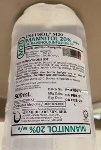
The ratio of solution mixed for patient to drink
400 ml Mannitol 20% mixed + 1100 ml water
(Total volume: 1500 ml)
Stoma Bag Preparation (if any)
180 ml solution of Mannitol + water injected into the stoma.
The ratio of solution mixed for stoma bag
30 ml Mannitol 20% mixed + 150 ml water
(Total volume: 180 ml)
Purpose of Mannitol + Water
- Hyperosmolar characteristic of Mannitol draws fluid into the bowel.
- Provides biphasic improved MRI soft tissue contrast.
- low signal intensity on T1-weighted images.
- high signal intensity on T2-weighted images.
Patient Positioning
Patient lying supine (head first) on the MRI table.
Give the head phone for patient’s comfort.
Briefly explain the patient about the alarm ‘squeezy’ button.
Cover patient’s body with blanket to keep warm.
Region of Interest Positioning
Plug in body coil (Large) at the system.
Wrap the coil horizontally over the patients’ abdominal area.
Marking the top border of the body coil at the level of xiphoid process.
IV Buscopan
To promote inhibition of intestinal peristalsis.
1 ml Buscopan (20 mg/ml) must be injected via IV line to the patient
2 doses.
- First: before start scanning.
- Second: During giving contrast.
SCANNING PROTOCOL
Protocol Overview
Plain
- haste_localizer
- t2_haste_cor_mbh
- t2_haste_tra_mbh
- t2_haste_fs_cor_mbh
- t1_vibe_fs_cor_bh
- t1_vibe_fs_tra_bh
- t2_trufi_cor_p2_bh
- t2_trufi_tra_p2_bh
Contrast
- t1_vibe_cor_bh_ce
- t1_vibe_tra_bh_ce
- ep2d_diff_b50_400_800_tra_p2_trig
—————————-
HASTE: half-Fourier acquired single-shot turbo spin echo
VIBE: volumetric interpolated breath-hold examination
TRUFI: true fast imaging with steady-state free precession
haste_localizer
Launch the scanning
Step 1 – haste localizer window will pop-up.
t2_haste_cor_mbh
3-axis haste images will apear in exam card.
Adjust the scanning box and parameters accordingly to create proper coronal image. (cover the small bowel)
mbh: multiple breath hold instruction will be given through out the scanning (2/3-times).
Right-click –>Edit Propertise –> Voice Command. Choose malay inhale and nafas biasa.
Pause: to create rest between instruction.
t2_haste_tra_mbh
3-axis haste images will apear in exam card.
Adjust the scanning box and parameters accordingly to create proper axial image. (cover the small bowel)
mbh: multiple breath hold instruction will be given through out the scanning.
The breath hold command need to be done manually (3-times).
Use F12 key to start each scan or click ‘PLAY’ button.
t2_haste_fs_cor_mbh
Copy t2_haste_cor_mbh protocol
Click Contrast.
Select Fat Saturation on
Tick Restore Magnitude on
t1_vibe_fs_cor_bh
3-axis images will apear in exam card.
Adjust the scanning box and parameters accordingly to create proper coronal image. (cover the small bowel)
bh: breath hold instruction will be given through out the scanning.
The breath hold command need to be done manually (1-time).
Please alert that the preset scanning will immediately start after user click Apply. To change, need to switch the Voice Command mode from No Command to Manual.
Use F12 key to start each scan or click ‘PLAY’ button.
t1_vibe_fs_tra_bh
Copy t1_vibe_fs_cor_bh protocol
Change the orientation from coronal to axial.
Adjust the slice per slab to cover the whole small bowel region.
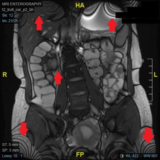
t2_trufi_cor_p2_bh
Drag the protocol from SIEMENS protocol card.
Adjust the scanning box and parameters to cover the small bowel region.
Use manual breath hold voice command.
Click Apply.
Use F12 key to start each scan or click ‘PLAY’ button.
The scanning will split into several scans (6 scans) with a rest in between scanning.
t2_trufi_tra_p2_bh
Drag the protocol from SIEMENS protocol card.
Adjust the scanning box and parameters to cover the small bowel region.
Use manual breath hold voice command.
Click Apply.
Use F12 key to start each scan or click ‘PLAY’ button.
The scanning will split into several scans (6 scans) with a rest in between scanning.
Moire Fringes / Zebra Artifact
An interference artifact that appear in the image when Gradient Echo is used with more than 1 coil.
Is a combination of aliasing artifact and field in-homogeneity
Due to superimposition of images with different phases from one side of the body to another.
These cause add and cancel signal.
How to Reduce Aliasing / Moire Fringes / Zebra Artifact?
Frequency Encoding Direction
- No frequency wrap (signal filter)
- Frequency Oversampling
Phase Encoding Direction
- Increase FOV
- Phase Oversampling
- Switch phase and frequency encoding direction
Only use surface coil (disable body coil)
3D
- Use z-gradient pulse during RF
Post Contrast
t1_vibe_cor_bh_ce
Open T1_vibe_fs_cor_bh protocol.
Copy slice and saturation region from t1_vibe_fs_cor_bh pre contrast.
Click Contrast.
Switch off fat saturation and restore magnitude to create non-fatsat protocol.
t1_vibe_tra_bh_ce
Repeat t1_vibe_tra_bh non-contrast protocol
Click Contrast.
Switch off fat saturation and restore magnitud to create non-fatsat protocol.
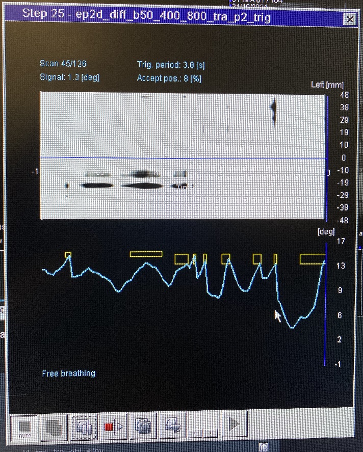
ep2d_diff_b50_400_800_tra_p2_trig
Trig: Breathing trigger
Can use 2 methods:
- Liver dome scout
- Can put the green box between the lung and liver. Small circle on the diaphragm.
- Phase scout
- Using a breathing triggering device (connect with ECG device). Put tightly between patient’s body and coil.
To use Phase scout:
- Click Physio –> PACE
- Resp. control: Trigger
- Scout type: Phase scout
- Signal1: none
To separate B value images
- Contrast –> Dynamic –> Multiple series: Each B value