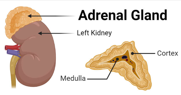
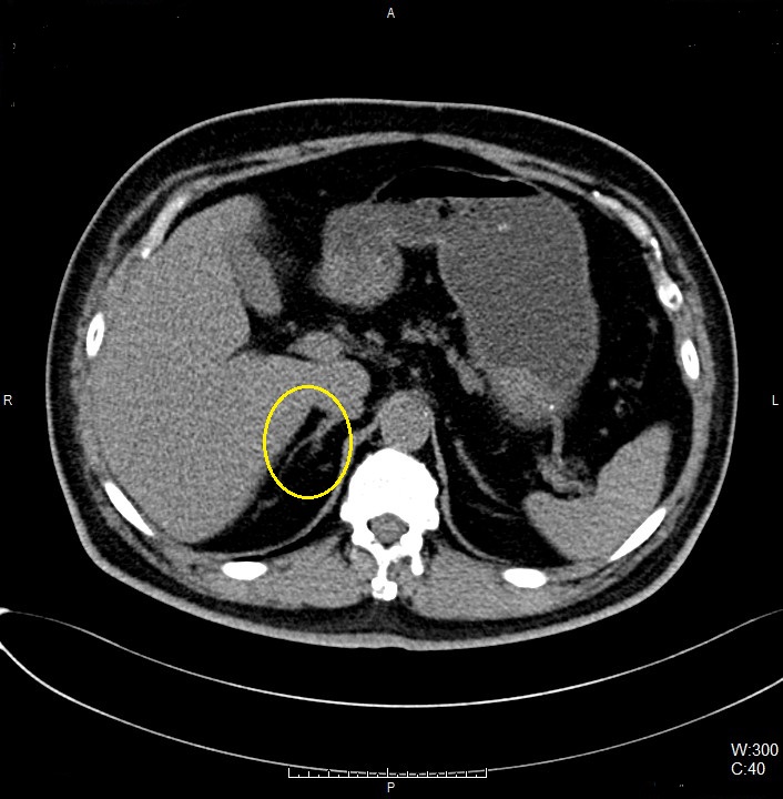
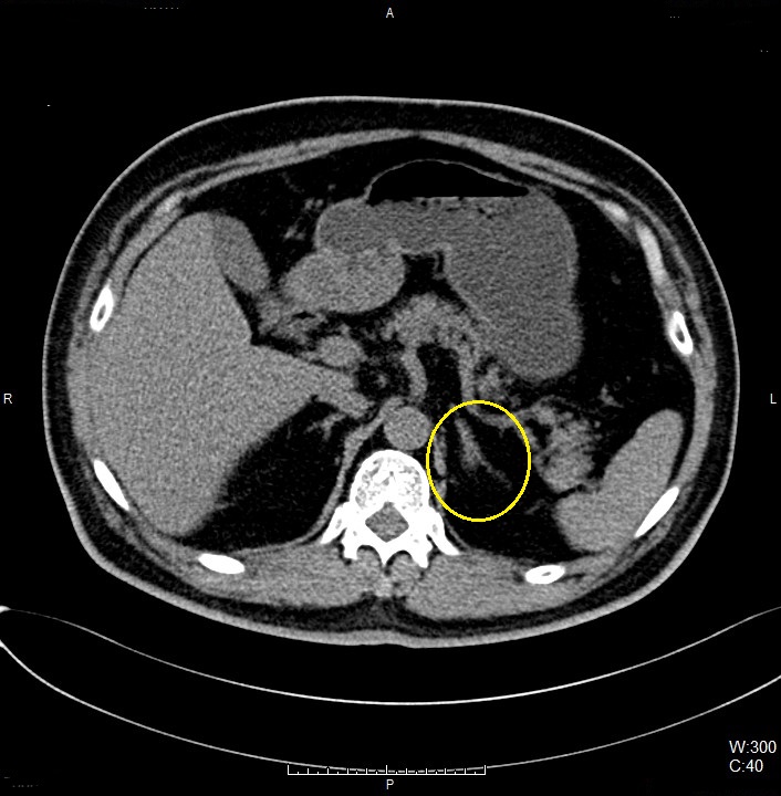
CT ADRENAL 3 PHASE Created June 28, 2021 Author Nabilah Nawi Category CT Scan Protocols Anatomy of Adrenal Gland Image taken from Microbe Notes Appearance of Adrenal Gland in Axial CT Image Right Adrenal Gland Left Adrenal Gland Protocol Structure 00_Liver3Phase (Adult) Topogram Non Contrast (full abdomen) Contrast Adrenal (cover adrenal and kidney) Portal Venous Phase (full abdomen) Delayed 15 Minutes (cover adrenal and kidney) Topogram Position the patient in head first supine position. Align the patient in Mid-Sagittal plane of the table. Position the transverse laser light beam at the level of patient’s nipple line to start the abdominal topogram. Topogram Parameters Topogram length: 512 cm Slice: 0.6 mm Scanning direction: Craniocoudal Tube position: Top Stop the topogram scanning when the scanning reach / pass over the inferior ischial ramus. Non Contrast Plan the Scan FOV (SFOV) box at topogram image Set the top line at the level of mid heart Set the bottom line at the level of inferior ischial ramus. Ensure the lateral line to cover patient’s body outline. Remind the patient before scanning as the breathing instruction will be given. Scanning Parameters kVp: mAs: Tube Current Modulation (TCM) Scanning Direction: Craniocaudal Scan Delay: 4 s Slice: Image Comment: Pitch: Reconstruction of Non Contrast Contrast Type of contrast used: Non-ionic iodinated contrast media 300 I mg/mol Needle Placement Test: Flow Rate: 3.5 ml/sec Volume of Normal Saline: 15 ml Contrast Injection: Volume of Contrast Media: 100 ml (depend on patient’s body habitus) Volume of Normal Saline: 50 ml Method of injection: Contrast Injector Flow Rate: Contrast: 3.0 ml/sec Normal Saline: 3.0 ml/sec Post Contrast Scan Planning Adrenal Plan the Scan FOV (SFOV) box at topogram image Set the top line at the level of mid heart Set the bottom line at the level below the kidneys. Ensure the lateral line to cover patient’s body outline. Portal Venous Phase Plan the Scan FOV (SFOV) box at topogram image Set the top line at the level of mid heart Set the bottom line at the level of inferior ischial ramus. Ensure the lateral line to cover patient’s body outline. Remind the patient before scanning as the breathing instruction will be given. Examination Overview Reconstruction of Adrenal Reconstruction of Portal Venous Phase Delayed 15 Minutes Plan the Scan FOV (SFOV) box at topogram image Set the top line at the level of mid heart Set the bottom line at the level below the kidneys. Ensure the lateral line to cover patient’s body outline. Remind the patient before scanning as the breathing instruction will be given. Multiplanar Reconstruction (MPR) Coronal Portal Venous Phase Image Thickness: 3.0 mm Number of Image: 19 Coverage: Anterior to Posterior of abdomen Series of Images Send to PACS Topogram Non Contrast 5.0 B30f Non Contrast 1.0 B20f Adrenal 5.0 B30f Adrenal 1.0 B20f PVP 5.0 B30f PVP 1.0 B20f Delayed 15 Min 5.0 B30f Delayed 15 Min 1.0 B20f Patient Protocol COR ST PVP Was this article helpful? Yes No
Leave A Comment?